Session 11
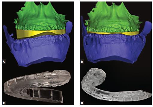
3D Virtual Imaging is one of the most significant tools for orthodontists to evaluate and record size and form of craniofacial structures. Orthodontists regularly use 2-dimensional (2D) static imaging procedures, but deepness of structures cannot be acquired and restricted with 2D imaging. Three-dimensional (3D) imaging has been developed in the early of 1990’s and has gained a prized place in dentistry, particularly in orthodontics. In 3D diagnostic imaging, a series of anatomical records is gathered using certain technical equipment, processed by a computer and later presented on a 2D monitor to present the illusion of deepness.
- Dental cone Beam computed tomography (CT)
- Cone Beam Imaging
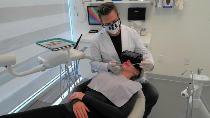
- Photography — extra oral and intraoral


No comments:
Post a Comment